
Use of underwater contactless ultrasonography to elucidate the internal anatomy and reproductive activity of manta and devil rays (Fam. Mobulidae)
May 2023
Niv Froman, Marie-Aude Genain, Guy M. W. Stevens & Gareth P. Pearce
Keywords: Anatomy • Elasmobranch • Mobulidae • Reproduction • Sexual Maturity • Ultrasonography
Summary: Traditional methods of studying the internal anatomy of fish require euthanasia and dissection, causing stress. A new waterproof, contactless, and portable ultrasonography equipment was developed and validated using manta and devil ray specimens from fish markets in Sri Lanka. Further validation was conducted on free-swimming reef manta rays, successfully identifying various internal structures. Ultrasonography proved to be a reliable and non-invasive method for determining sexual maturity and gestational status in these rays, offering a practical alternative to invasive techniques in studying anatomical changes in marine organisms.
Abstract
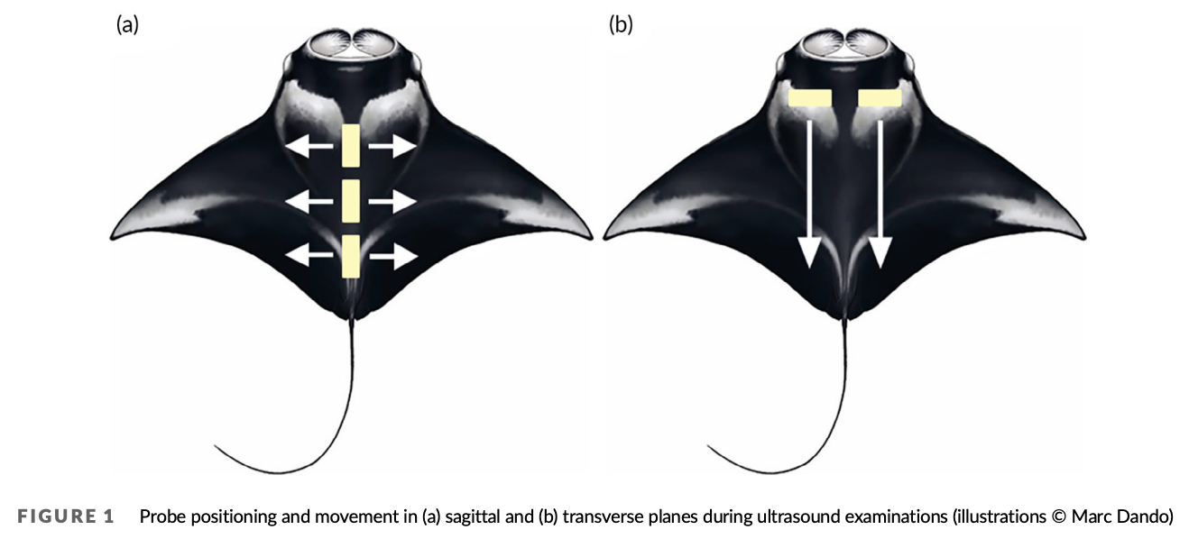
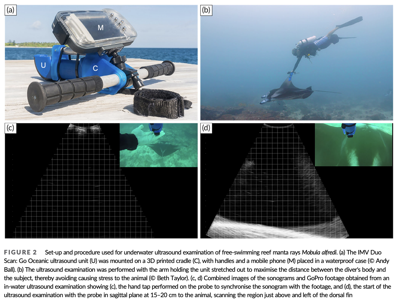
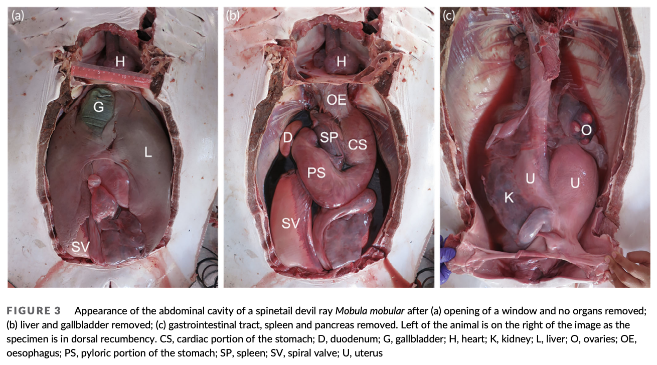
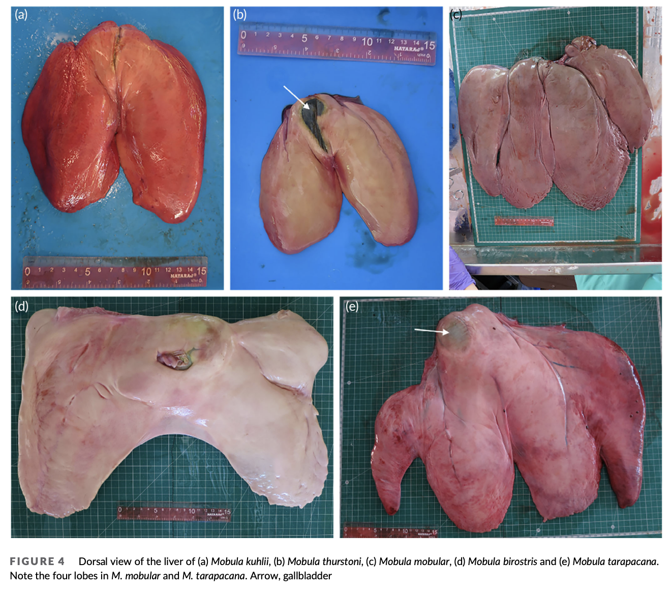
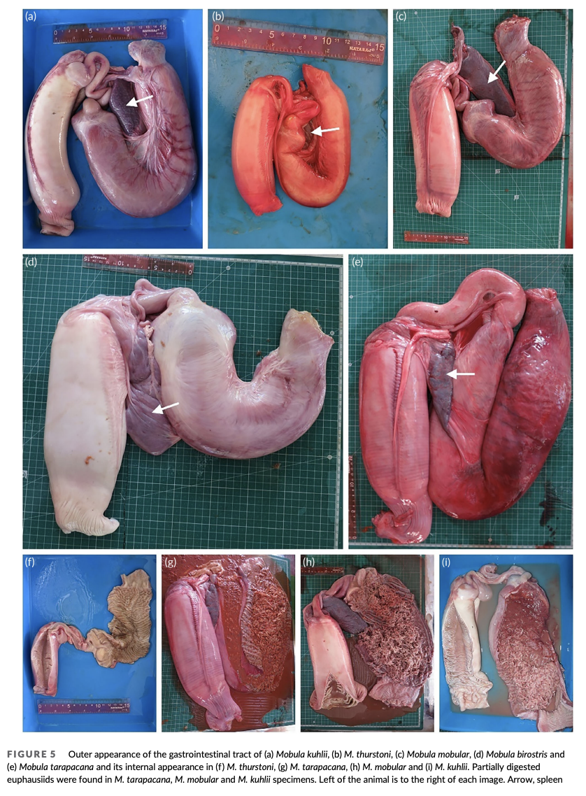
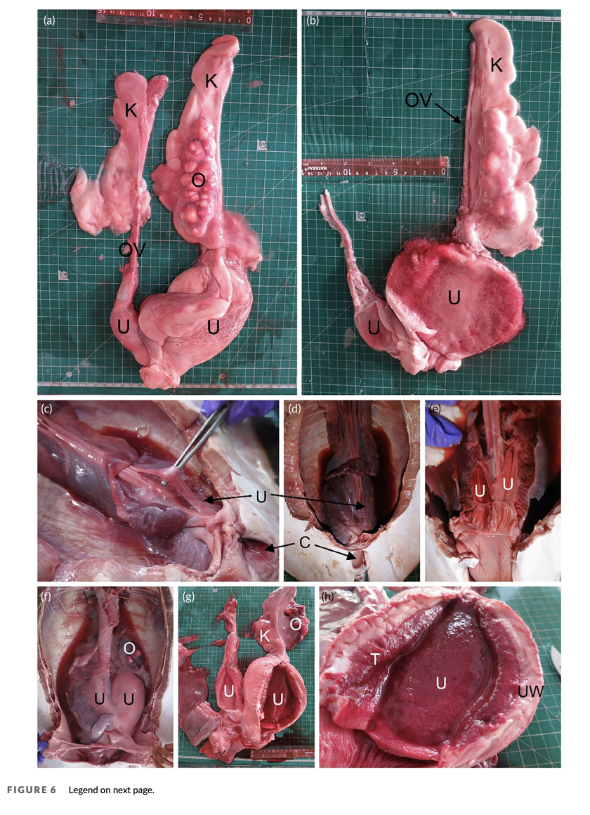
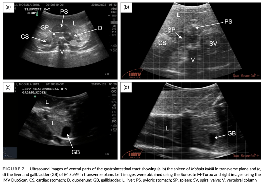
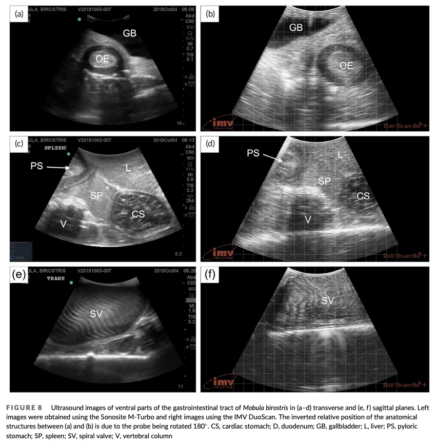
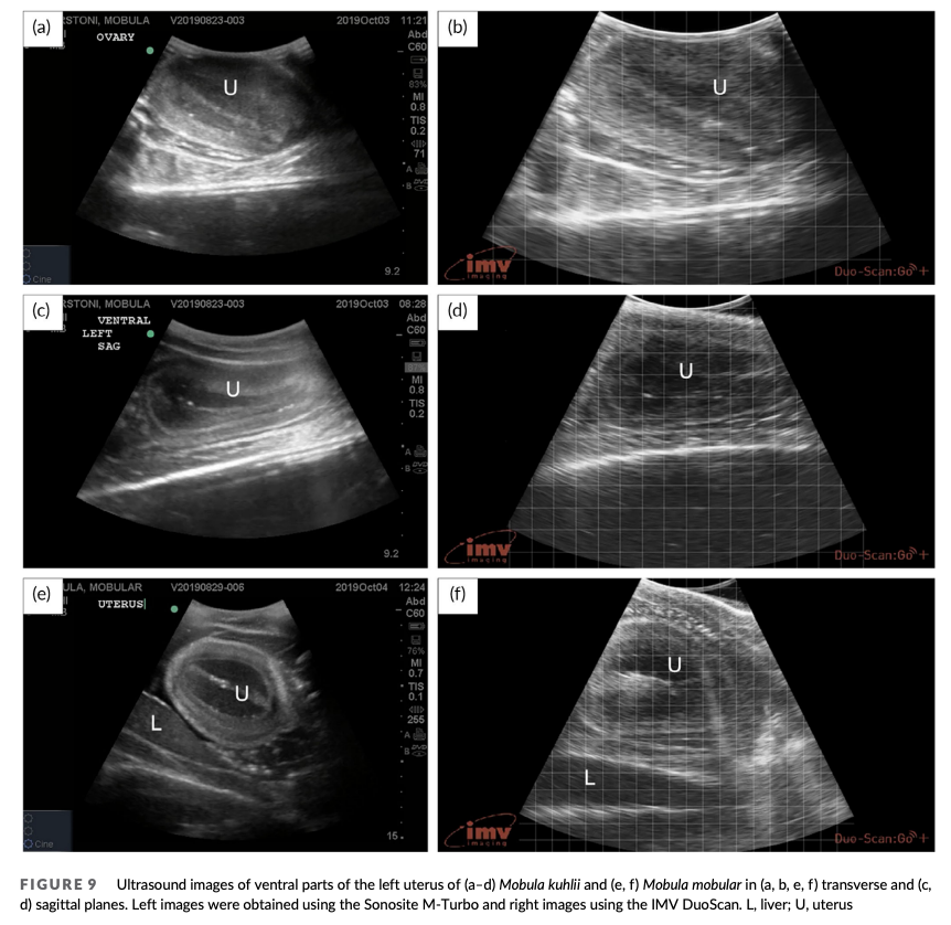
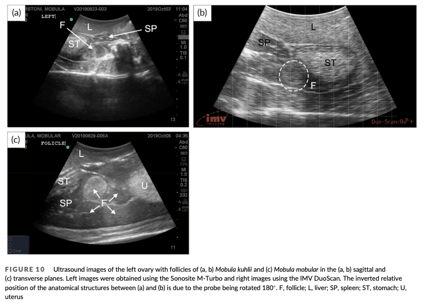
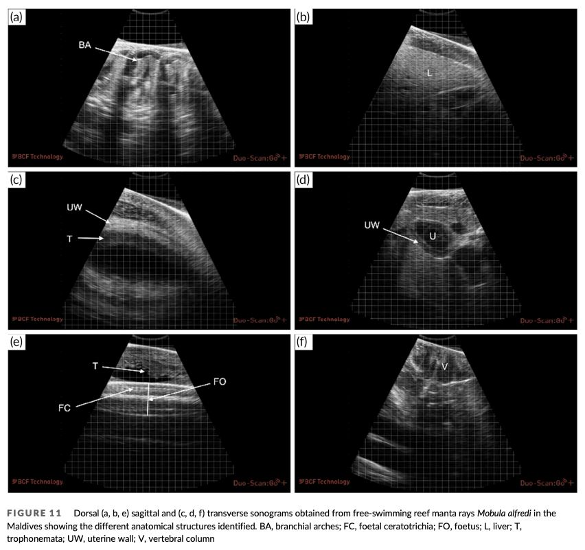
“The ability to visualise the internal anatomical structures of fish provides important information on their reproductive status and body condition and has made important contributions to many areas of fish biology. Obtaining information on the internal anatomy of fish has traditionally required euthanasia and dissection. Although ultrasonography is now increasingly used to study internal fish anatomy without the need for euthanasia, traditional techniques still require restraint and contact with the animal, both of which are known to cause stress. This has prompted the development of waterproof, contactless and portable equipment to allow ultrasonographic examinations to be carried out in free-swimming individuals, which also facilitates the application of this tool in wild populations of endangered species. This study reports the validation of this equipment using anatomical examinations of nine manta and devil ray (Mobulidae) specimens landed at fish markets in Sri Lanka. The species studied were Mobula kuhlii (n = 3), Mobula thurstoni (n = 1), Mobula mobular (n = 1), Mobula tarapacana (n = 1) and Mobula birostris (n = 3). The use of this equipment was further validated with ultrasonographic examinations in 55 free-swimming reef manta rays Mobula alfredi, which enabled maturity status to be quantified in 32 females. Structures successfully identified in free-swimming individuals were the liver, spleen, gallbladder, gastrointestinal tract, skeletal structures, developing follicles and uterus. The study demonstrated that ultrasonography provided a reliable method of determining both sexual maturity and gestational status in free-swimming M. alfredi. The methodology induced no detectable signs of disturbance to the animals involved and therefore offers a viable and practical alternative to invasive techniques currently used to study anatomical changes in both captive and wild marine organisms.”
Infographics

In The Press

Author Affiliations
The Manta Trust
Vetsonic Ltd
Flying Sharks
Manta Expeditions Ltd
University of Cambridge
Blue Resources Trust
Funded by
The Manta Trust
Vetsonic Ltd
Flying Sharks
Manta Expeditions Ltd
University of Cambridge
Six Senses Laamu